The role of the brain
The brain is an essential, complex organ that controls the functions and processes of the body. It receives input from the rest of the body, such as from the five senses, as well as autonomic (involuntary) input from the organs, and interprets this information.
The brain enables thoughts, emotions, motor skills and perceptions of sensations, and also regulates heart rate, blood pressure and breathing, the release of hormones, and the processing of sensory information, among other functions.
Anatomy: the main parts of the brain and their functions
The brain is made up of a large mass of nerve tissue and consists of many specialised areas that work together. It is not a muscle.
The main parts of the brain are the cerebrum, brainstem and cerebellum. It is surrounded by a layer of tissue called the meninges and sits inside the skull (cranium), which helps protect it from injury.
Cerebrum
The cerebrum is the largest part of the brain. The outermost layer of cells, which gives the brain its wrinkled surface, is called the cerebral cortex and consists of grey matter. The centre consists of white matter.
The cerebral cortex is made up of a left half and a right half (called hemispheres). The right hemisphere controls the left side of the body, while the left hemisphere controls the right side of the body. The left hemisphere is commonly the “dominant” one. Most right-handed people are left hemisphere dominant, while left-handed people are sometimes right hemisphere dominant. The dominant hemisphere is typically responsible for speech and language functions, while the non-dominant hemisphere is responsible for spatial awareness and processing sensory information.
Each hemisphere is divided into regions called “lobes” that work together. Each lobe plays an important role in specific functions, such as interpreting sensations from the body, processing sights, sounds and smells, thoughts, problem solving and planning, forming and storing memories, and controlling voluntary movement.
- Frontal lobes: These are the largest lobes and are located at the front part of the brain, right behind the forehead. The frontal lobes coordinate and manage voluntary movements, speech and thought, and behaviours such as motor skills, problem solving, judgment, emotions and personality.
- Parietal lobes: These lobes are located behind the frontal lobes, near the centre of the brain, and are involved in managing sensation and body position, and organising and interpreting sensory information from other parts of the brain. This part of the brain helps you understand your environment and your body’s state.
- Temporal lobes: These are located on either side of the head, level with the ears. They coordinate functions such as hearing, visual and verbal memory. This helps you understand language and recognise people and places.
- Occipital lobes: These are located at the back of the brain and enable you to note and interpret visual information, such as recognising colours, shapes and movement.
The two hemispheres of the brain communicate with each other through a large, C-shaped structure called the corpus callosum that consists of white matter and nerve pathways.

The main parts of the brain are the cerebrum, cerebellum and brain stem. The cerebrum consists of two hemispheres, each of which is divided into lobes: frontal lobes, parietal lobes, temporal lobes and occipital lobes.
Cerebellum
The cerebellum sits at the base and back of the brain, under the cerebrum. It is responsible for fine motor skills, such as coordination of smaller movements, and balance and posture.
Brain stem
The brain stem is located beneath the cerebrum and in front of the cerebellum. It is made up of three major parts: midbrain, pons and medulla oblongata. It connects the brain to the spinal cord, which runs down the neck and back, and is responsible for passing messages to various parts of the body and the cerebral cortex.
The brain stem controls the involuntary muscles in the body – those that work automatically, such as the heart and the digestive system – and the functions of the body needed for life, such as heart rate, blood pressure, sleep and breathing.
How does the brain work?
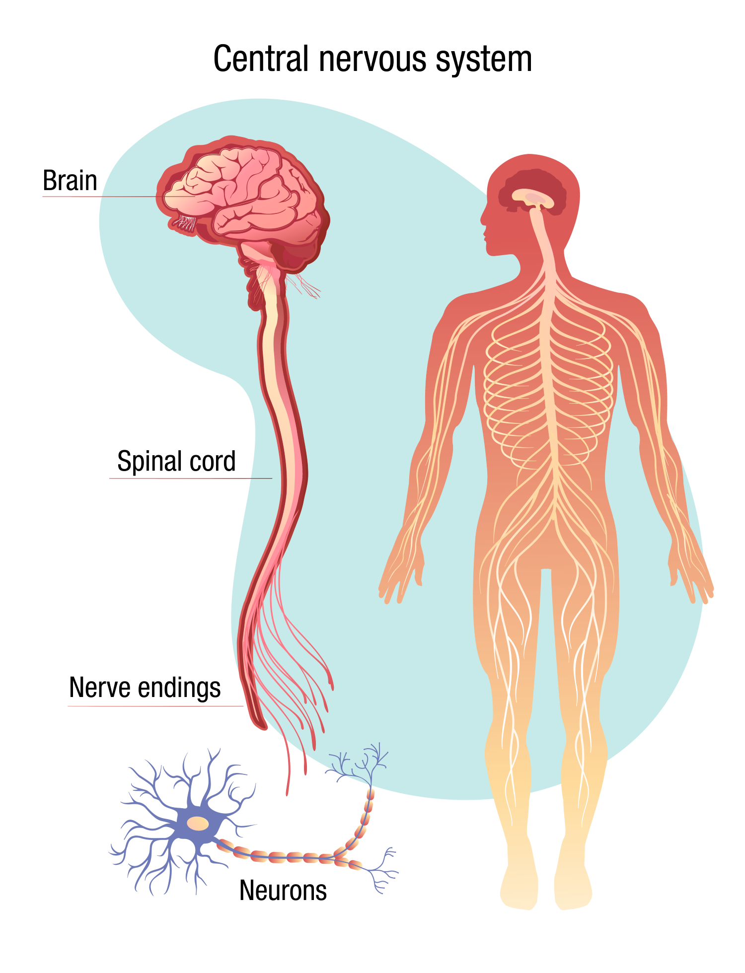
The brain is connected to the spinal cord; together they make up the central nervous system.
The central nervous system contains millions of neurons (nerve cells) that branch out to every organ and body part. Neurons connect and communicate with each other at junctions called synapses.
Through the central nervous system, the brain sends and receives signals throughout the body. These signals can be chemical or electrical, and different signals control different processes. The many specialised areas of the brain work together to interpret these signals.
The signals move through individual neurons as tiny electrical charges. When a charge reaches a synapse, it may trigger the release of chemicals called neurotransmitters. Neurotransmitters act as chemical messengers, carrying messages from one neuron to the next target cell.
When learning something new, messages repeatedly travel from neuron to neuron. Eventually, the brain starts to create connections (pathways) between the neurons, which makes the new activity easier. Over time, patterns are created in signal type and strength.

Connection between the brain and the heart
The brain needs a constant supply of oxygen-rich blood to function properly. This blood is pumped to the brain by the heart through two sets of blood vessels: the vertebral arteries and the carotid arteries. The external carotid arteries run up the sides of the neck and are one location where the pulse can be felt.
The brain is also connected to the heart, and other organs, through the vagus nerves. These nerves allow the brain to receive information about how the heart is functioning and send commands to the heart, such as how quickly it should beat.
The brain and the heart are in constant communication to keep the body functioning.
Connection between the brain and blood pressure
The brain is supplied with blood and oxygen through arteries. High blood pressure, which is when the force of the blood against the artery walls is higher than normal for an extended period, can damage the arteries. This can cause them to narrow, rupture or leak, or cause the formation of blood clots.
When blood flow to the brain is blocked and the brain does not get enough oxygen and nutrients, this can cause a stroke. Brain cells will start dying until blood flow is restored.
Research by the Heart Research Institute (HRI) is investigating how the brain controls breathing and blood pressure, and the relationship between blood pressure and obstructive sleep apnoea.
Brain conditions
There are many types of disorders and conditions, varying in severity, that can affect the brain.
Some of these may be due to inherited conditions or developmental disorders, while others may progressively develop with age, such as Alzheimer’s disease and dementia. Recent research by HRI has also found a strong link between dementia and atrial fibrillation, an irregular heartbeat condition.
Some brain conditions may be the result of an injury, such as a blow to the head.
Other conditions in the body, such as cardiovascular disease and blood clots, can cause a stroke that can lead to brain damage.
If you or someone you know displays symptoms of a brain condition such as stroke or aneurysm, seek medical help immediately.
Symptoms of blood clot in the brain
If a blood clot occurs in an artery to the brain, stopping the brain from getting the blood and oxygen supply that it needs, this can cause a stroke. Brain tissue starts dying without a constant supply of oxygen, so it is critical to treat the stroke as soon as possible. The longer a stroke remains untreated, the greater the chance that brain cells will die, resulting in permanent stroke-related brain damage.
These debilitating effects can include weakness on one side of the body, difficulty controlling movements, personality or behaviour changes, or problems speaking and understanding.
Symptoms of blood clot in the brain depend on which part of the brain the clot occurs in, but they are the same as the symptoms of a stroke. These can include:
- numbness or weakness in the arm, face or leg (especially on one side of the body)
- slurred speech or trouble speaking or understanding others
- dizziness
- sudden, severe headache
- vision problems.
Brain aneurysm symptoms
An aneurysm in the brain (cerebral aneurysm) is when an abnormal bulge or swelling occurs in the wall of a blood vessel in the brain. Over time, this bulge typically grows larger and the artery walls become weaker.
If an aneurysm ruptures and bleeds, blood leaks into the area around the brain (subarachnoid haemorrhage). This haemorrhage can also directly damage the brain – called a haemorrhagic stroke.
Symptoms when a brain aneurysm ruptures include:
- sudden, severe headache
- pain and stiffness in the neck
- drowsiness
- paralysis
- seizures
- impaired speech and visual problems.
A brain aneurysm may have no symptoms until it is either very large or it ruptures.
The cause of a brain aneurysm is not always known. High blood pressure over the long term can damage and weaken blood vessels, making them more likely to rupture. Atherosclerosis, the main underlying cause of cardiovascular disease, can also weaken blood vessel walls when there is a build-up of atherosclerotic plaque.
How is HRI working to help the brain?
HRI is tackling cardiovascular disease and its effects on organs such as the heart and brain from a broad range of research angles.
The mission of our Heart Rhythm and Stroke Prevention Group is to prevent as many strokes as possible – and stroke-related brain damage – through early detection of the abnormal heartbeat condition atrial fibrillation (AF), which is linked to one in three strokes. With a clinical implementation focus, the Group is exploring novel strategies using eHealth tools and patient self-screening to detect unknown silent AF. People with AF are also at increased risk of dementia. Earlier diagnosis of AF could be considered a strategy to prevent or delay dementia and stroke.
The Microvascular Research Group is investigating how sepsis, a life-threatening complication of an infection, impacts the normal function of the arteries, with the goal of identifying a treatment target to reverse the drop in blood pressure seen in septic shock. Sepsis can have a devastating impact on the cardiovascular system and cause dysfunctions in major organs of the body, including the brain, lungs, kidneys and liver.
